
Learn what dense breasts are, why they can raise cancer risk, and how research is closing the screening gap.
Key Takeaways
- Nearly half of women over 40 have dense breasts.
- Dense breasts are largely related to genetics, but other factors, like age and weight, can play a role.
- Individuals with dense breasts have a higher risk of breast cancer, and dense tissue can make it harder to detect on mammograms.
- BCRF researchers are studying new screening methods for women with dense breasts.
Dense breasts are a growing focus of breast cancer research. Around half of women over 40 have them, and they are associated with a higher risk of developing breast cancer. But what exactly are they?
Ahead, discover important things to know, like what causes dense breast tissue—and should you worry if you have dense breasts?
What are dense breasts?
Breasts contain three types of tissue. Fibrous (also called connective) tissue keeps the breast in place; glandular tissue, which includes lobules and ducts, produces and transports breastmilk. Together, these two tissues are called fibroglandular tissue. The third type is fatty tissue, which surrounds the other tissues and helps create the size and shape of the breast.
Having dense breasts means you have more fibroglandular tissue than fatty tissue—and it’s completely normal. In fact, dense breast tissue occurs in nearly half of women over the age of 40.
What are the types of dense breasts?
Breast density can range from entirely fatty to extremely dense. On a mammography report, radiologists classify the density of a woman’s breast into one of four categories:
A: Entirely or almost entirely fatty. Breasts in this category are almost entirely made up of fatty breast tissue. This means that they contain very little fibroglandular (dense) tissue. This occurs in about 10% of women.
B: Scattered areas of dense breast tissue (a.k.a. fibroglandular density). This category does not mean a woman has “dense breasts” and it occurs in about 40%of women.
C: Heterogeneously dense. These are considered “dense breasts.” Here, there are large areas of dense breast tissue throughout the breasts. This occurs in about 40%of women.
D: Extremely dense. Breasts in this category have extremely dense breast tissue, which means that most of the breast is made of fibroglandular tissue. About 10% of women have extremely dense breasts.
How do you know you have dense breast tissue?
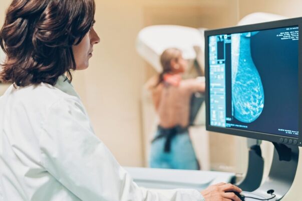
Breast tissue density is detected through a mammogram. Fibroglandular (dense) tissue shows up as white, while fatty tissue is gray or black. So, the whiter the mammogram, the more dense tissue there is.
What causes dense breast tissue?
Dense breasts are not rare— they’re the reality for nearly half of all women over age 40. Whether you have dense breast tissue largely depends on your genetics. However, other factors include:
- Being younger than 40. Breast density is higher in women under the age of 40 and generally decreases over time. Dense breasts occur in 40% of women in their 50s and 25% of women aged 60.
- Being pregnant or breastfeeding.
- Taking hormone replacement therapy.
- Having a lower body weight. Less body fat means less fatty tissue in the breasts.
Race and ethnicity can also play a role. Asian women and Black women are more likely to have dense breast tissue than Hispanic women or white women—groups who, in different ways, already face greater risks and/or disparities in breast cancer outcomes.
Are women with dense breasts more likely to have cancer?
Women with dense breasts face a 15 to 20%increased risk of developing breast cancer compared to the national average, according to the American Cancer Society (ACS). Scientists are still working to uncover why that is. Since breast cancer often develops in glandular tissue, having higher amounts of it may simply create more opportunities for abnormal cells to grow and multiply.
It’s worth noting that breast cancer patients with dense breasts do not face a higher risk of death than individuals with non-dense breasts, per the Centers for Disease Control and Prevention (CDC).
Does dense breast tissue make mammograms harder to read?
Dense breast tissue can make it more difficult for radiologists to spot cancer on a standard mammogram (also called a 2D mammogram). While fatty tissue appears almost black on a mammogram, dense glandular and fibrous tissue looks light gray or white, like breast masses do. That overlap can hide tumors in the image and increase the risk that a potentially cancerous growth will go undetected. This also contributes to higher rates of late breast cancer diagnoses among women with extremely dense breasts, according to a 2022 report in Springer.
However, while standard mammograms aren’t perfect, they still can show most breast cancers,including those in dense breasts. As of 2024, the U.S. Food & Drug Administration (FDA) requires all mammogram facilities in the U.S. to notify patients of their breast density in their report. That means you’ll have an updated status on your breast density after every mammogram and you can work with your doctor to decide whether additional screening measures might be right for you.
Is there treatment for dense breast tissue?
Dense breast tissue is not a problem in and of itself and cannot be reduced. However, women with dense breasts can access more advanced screening tools to increase the likelihood of detecting cancer.
More than 90% of all screening facilities in the U.S. now offer 3D mammograms, according to FDA data from March 2025. Also known as digital breast tomosynthesis, a 3D mammogram uses low-dose X-rays to take multiple images of the breast in an arc-like pattern. Software then reconstructs those images into thin “slices,” which radiologists can review one by one to check for abnormal growths.
Research has found that 3D mammograms, which are already part of standard breast cancer screening for many women, improve detection rates in women with heterogeneously dense breasts. 3D imaging, however, can still miss cancers within very dense tissue.
Experts have yet to determine which secondary cancer screening tests, if any, should be standard for women with dense breasts. Healthcare professionals may recommend whole breast ultrasounds, which use sound waves to help differentiate between likely benign fluid-filled masses and potentially cancerous solid masses. Another common option is breast magnetic resonance imaging (MRI), which uses magnets and radio waves to form an image of the breast. This type of screening can help a radiologist detect lesions hidden within breast tissue more accurately than mammograms or ultrasounds, but it can be cost prohibitive.
These tools are more sensitive, which means they can highlight cancers that a mammogram might miss. The flip side is that they can also flag areas that aren’t cancer at all (false positives), leading to extra tests, biopsies, and worry for patients.
Are there new tools to screen women with dense breasts for cancer?
As those with dense breasts face both a higher risk of cancer and less precise screening, BCRF researchers are studying new tools for earlier and more accurate detection. They’ve already found that abbreviated breast MRIs can detect invasive breast cancer more effectively than 3D mammography in women with dense breasts. They’ve also discovered that Molecular Breast Imaging (MBI) can help doctors see rapidly dividing cells that may be cancerous.
BCRF researchers are also evaluating contrast-enhanced mammography (CEM) to see if it is more effective than 3D mammography at detecting cancer in women with dense breasts. The results of these trials could open the door to new screening standards for women with dense breasts.
Research into dense breasts can help inform the next generation of breast cancer screening and ensure that women with dense breasts get clearer answers about their health—ultimately bringing us closer to our goal of zero deaths from breast cancer.

