

Dense Breast Tissue: What It Means and What to Know
BCRF shares what it means to have dense breasts and how researchers are striving to improve screening for women who have them
Dense breasts are associated with an elevated risk of developing breast cancer, in part because dense breast tissue can make it difficult for doctors to see abnormal growths in mammograms that may be cancer.
So, what does it mean to have dense breast tissue, and why is it important to know if you have it? BCRF dives into this topic and highlights research focused on improving breast cancer detection in women who have dense breasts.
What is dense breast tissue?
Breasts are made up of different types of tissue: Fibrous or connective tissue that holds the breast in place; glandular tissue, which includes lobules and ducts that produce and transport milk; and fatty tissue that fills the space between fibrous and glandular tissue and helps give breasts their size and shape. Breast density is a measure of how much fibrous and glandular tissue (together referred to as fibroglandular tissue) there is in the breast relative to fatty tissue. If you have higher breast density, it means that your breasts are comprised of more fibroglandular tissue compared to fatty tissue.
Dense breast tissue is normal and occurs in nearly half of women over the age of 40. On average, breast density is higher in women under the age of 40 and tends to decrease as women get older; 40 percent of women in their 50s and 25 percent of women aged 60 and over have dense breasts.
Asian women are more likely to have dense breast tissue than women of other races. And a 2023 study of nearly 900,000 women in the Breast Cancer Surveillance Consortium found that Black women are more likely to have dense breasts than Hispanic or white women after adjusting for age, menopausal status, and body mass index.
According to the Centers for Disease Control, women are more likely to have dense breast tissue if they are pregnant or breastfeeding, take hormone replacement therapy, or have a lower body weight.
How is dense breast tissue detected?
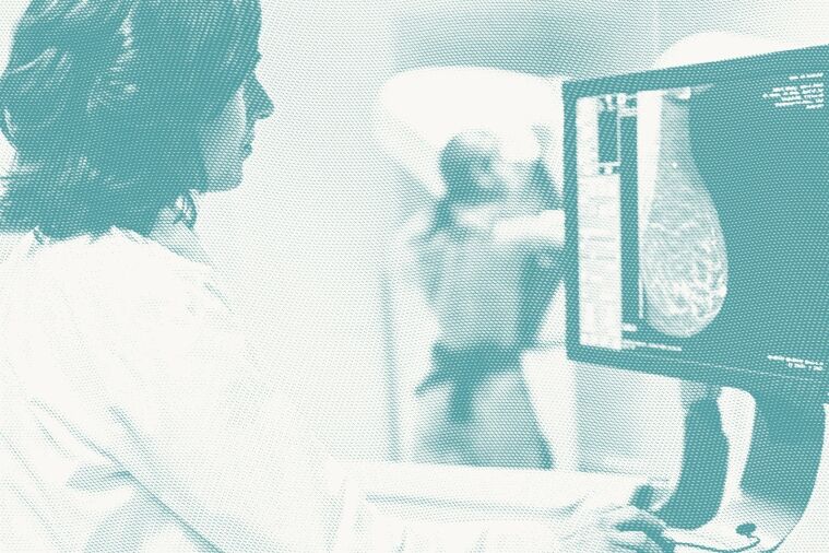
A radiologist reviews a mammogram and assesses the proportion of white, gray, and black areas on the image to detect dense breast tissue. Fibroglandular tissue appears white on a mammogram, so the greater proportion of white there is on the image, the denser the breasts. Breasts that feel firm to the touch do not necessarily indicate a higher breast density, so dense breasts cannot be detected during a physical examination.
As of September 2024, the FDA requires that all mammogram facilities nationwide include a breast density statement in a patient’s report. That statement designates the patient’s breasts as dense or not dense and describes how breast density can influence the interpretation of a mammogram. Prior to this rule, reporting standards for dense breast tissue varied from state to state, and several did not require any patient notification.
Why does dense breast tissue matter?
Breast density is just one factor associated with an elevated breast cancer risk. Researchers are still trying to understand the underlying links between dense breasts and cancer. Breast cancer is known to develop in glandular tissue, so having a greater amount of it may simply provide more opportunity for abnormal cells to grow.
Higher breast density can also make it more challenging for radiologists to see abnormal growths since they both appear white on a mammogram. Dense breast tissue can effectively “cloud” the mammogram and increase the likelihood that a potentially cancerous growth or tumor gets missed.
BCRF investigator Dr. Graham Colditz is one breast cancer researcher who is interested in developing better methods to predict breast cancer in women with dense breast tissue. He and his team have shown that while breast density generally decreases with age, the rate of breast density decrease was significantly slower in a breast that was later diagnosed with cancer compared to breasts that remained cancer-free. They have collected and analyzed mammogram images from 10,000 women to develop a model to predict cancer development. In a related study, Dr. Colditz and his team have also shown that having a family history of breast cancer was associated with having dense breasts in premenopausal women.
How is dense breast tissue categorized?
Doctors use a system developed by the American College of Radiology (ACR) called Breast Imaging Reporting and Data System (BI-RADS) to interpret and report standardized mammogram findings. It is subjective in nature, so doctors are increasingly using automated software to assist in the classification process. BI-RADS classifies dense breasts into four categories:
Category A: fatty breast tissue
Category A breasts are composed almost entirely of fatty breast tissue, with very little fibroglandular (dense) tissue present. On a mammogram, this type of breast appears mostly dark gray or black, since fat shows up as darker areas on breast imaging. Small amounts of dense tissue may still appear as light gray or white. Approximately 10 percent of women fall into this category.
Category B: scattered areas of dense breast tissue
Category B is defined by breasts with scattered areas of fibroglandular (dense) tissue. On a mammogram, this presents as a mix of light and dark gray or black areas. Because both dense and non-dense tissue are visible, some regions of the breast may be more difficult to evaluate, and tumors can be obscured in areas that appear dense. About 40 percent of women fall into Category B.
Category C: heterogeneously dense breasts
Category C refers to heterogeneously dense breast tissue, where there are large areas of fibroglandular tissue present throughout the breast. On a mammogram, these breasts appear predominantly light gray or white because dense tissue shows up as white on breast imaging. This can make it more difficult to distinguish certain findings since both dense tissue and some abnormalities appear similar on a mammogram. Approximately 40 percent of women fall into this category.
Category D: extremely dense breasts
Category D is defined by extremely dense breast tissue, where most of the breast is composed of fibroglandular tissue. On a mammogram, these breasts appear almost entirely white due to the high concentration of dense tissue. This high density can make it harder to see certain findings on a mammogram because dense tissue and potential abnormalities may appear similar. Around 10 percent of women fall into Category D.
Women in categories C and D are considered to have dense breasts.
Screening options for women with dense breast tissue
Dense breast tissue can make standard mammograms more challenging to interpret because both dense tissue and potential abnormalities appear white on breast imaging. This overlap can sometimes make it harder for radiologists to distinguish between normal tissue and areas that may require further evaluation.
There is currently no single agreed-upon approach for supplemental screening in women with dense breasts. Recommendations vary depending on breast density category, overall risk factors, and clinical judgement made by a radiologist in consultation with a patient’s doctor.
For women with heterogeneously dense (Category C) or extremely dense (Category D) breast tissue, these additional screening methods may sometimes be considered:
3D mammography (tomosynthesis)
Today, many women receive 3D mammograms (also called tomosynthesis) as part of routine screening. While tomosynthesis does not create a true three-dimensional image, it provides a clearer view of internal breast structures compared with standard 2D mammography.
Like standard 2D mammography, 3D mammography uses X-rays to create images of the breast. However, it captures multiple images from different angles and uses software to reconstruct the breast into thin image “slices.” These slices allow radiologists to view the breast in greater detail and reduce the impact of overlapping tissue.
3D mammography has been shown to improve breast cancer detection in women with heterogeneously dense breasts (Category C), although results are more mixed in women with extremely dense breasts (Category D), because the more white area there is on the image, the more hidden a growth can be.
Ultrasound screening
Whole breast ultrasound uses high-frequency sound waves to create images of the breast. It is sometimes used as a supplemental screening tool following a 2D or 3D mammogram.
Ultrasound does not use ionizing radiation and is generally well tolerated. It can help distinguish between fluid-filled cysts, which are typically benign, and solid masses, which may require further evaluation to rule out cancer.
Breast MRI
Breast magnetic resonance imaging (MRI) is a noninvasive imaging test that uses strong magnets and radio waves to produce detailed images of the breast. A gadolinium-based contrast agent is injected intravenously to improve visualization of breast tissue and blood flow.
MRI is highly sensitive and can detect lesions that may not be visible on mammography or ultrasound. However, it is not always used as a first-line screening tool because it can be costly and may lead to findings that require additional follow-up even when they are not cancer. These findings are often referred to as false positives.
Contrast-enhanced mammography (CEM)
Contrast-enhanced mammography (CEM), also known as contrast-enhanced spectral mammography (CESM) or contrast-enhanced digital mammography (CEDM), is an emerging imaging technique that combines traditional mammography with an intravenous iodine-based contrast agent. Cancer cells attract more of the iodine-containing contrast agent compared to normal cells, so it is easier to identify cancerous tissue in CEM than through standard 2D or 3D mammography.
CEM is currently being studied as part of ongoing research into improving breast cancer detection, particularly in women with dense breast tissue. It is not yet universally used as a standard screening tool but is part of evolving approaches aimed at improving diagnostic accuracy.
Emerging research on dense breast tissue and cancer detection
BCRF investigators are testing new screening technologies to better screen dense breast tissue while reducing unnecessary follow-up procedures.
Dr. Wendie Berg is examining cancer detection and false positive rates in 3D mammography screening compared to CEM. Dr. Berg, who is also the Chief Scientific Advisor to DenseBreast-info.org, is conducting several studies of contrast-enhanced mammography in women with a personal history of breast cancer. She has completed enrollment to the first trial, Tomosynthesis or Contrast Enhanced Mammography (TOCEM), which is examining cancer detection rates in women screened annually via 3D mammography and CEM for a period of four years. The second study, Biennial CEM with Annual Tomosynthesis (BI-TOCEM) is open to enrollment. It is evaluating every-other-year supplemental screening with CEM in addition to annual 3D mammography for a period of five years. Dr. Berg and her team have observed that cancer detection rates using CEM are similar to rates from MRI screening but with fewer callbacks and false positives. CEM also has lower costs and shorter examination times compared to MRI screening methods, and it is an alternative choice for patients with claustrophobia or metallic implants.
Dr. Christopher Comstock is also evaluating the use of CEM as a breast cancer screening option for women with dense breast tissue. He is the principal investigator for the Contrast-Enhanced Mammography Imaging Screening Trial (CMIST), supported by BCRF in collaboration with ACR and GE Healthcare. The trial has opened at 10 sites that are currently enrolling patients. Ongoing analysis will help determine if CEM improves cancer detection and reduces false positives in women with dense breasts.
Two other new screening techniques may be beneficial for women with dense breast tissue:
- An abbreviated breast MRI—which uses the same technology as traditional MRI but takes fewer images over a shorter time period—has been shown to have better invasive breast cancer detection rates compared with tomosynthesis among women with dense breasts. In addition to shorter screening times, abbreviated breast MRIs are less costly.
- Molecular Breast Imaging (MBI), also known as scintimammography or breast-specific gamma imaging, uses radioactive material injected into the blood that accumulates more often in cancer cells than in normal cells. A gamma ray camera detects the gamma rays the radioactive tracer emits. This technique does not provide the same anatomical information as a standard mammogram or ultrasound, but it allows doctors to visualize specific cells as they function within breast tissue—for example, rapidly dividing cells that may indicate cancer.
One size does not fit all
There are many considerations when it comes to breast cancer screening and care. Having dense breasts is one of them. Knowing if you have dense breasts is important when considering screening options. You can ask your doctor if you have dense breast tissue and, if so, discuss how you should be screened for breast cancer.
Support research in early detection
Support the research that’s helping to improve breast cancer screening and detection for women with dense breasts. Your gift to BCRF helps fund scientists working to advance imaging technologies and improve outcomes through earlier and more accurate diagnosis. Donate, start a fundraiser, or explore more ways to give today.
Selected References 
About dense breasts. (2024, September 11). Breast Cancer. https://www.cdc.gov/breast-cancer/about/dense-breasts.html?CDC_AAref_Val=https://www.cdc.gov/cancer/breast/basic_info/dense-breasts.htm#
Brand, J. S., Czene, K., Shepherd, J. A., Leifland, K., Heddson, B., Sundbom, A., Eriksson, M., Li, J., Humphreys, K., & Hall, P. (2014). Automated Measurement of Volumetric Mammographic Density: a tool for widespread breast cancer risk assessment. Cancer Epidemiology Biomarkers & Prevention, 23(9), 1764–1772. https://doi.org/10.1158/1055-9965.epi-13-1219
Del Carmen, M. G., Halpern, E. F., Kopans, D. B., Moy, B., Moore, R. H., Goss, P. E., & Hughes, K. S. (2007). Mammographic breast density and race. American Journal of Roentgenology, 188(4), 1147–1150. https://doi.org/10.2214/ajr.06.0619
Dense breast tissue | Breast Density and Mammogram reports. (n.d.). American Cancer Society. https://www.cancer.org/cancer/types/breast-cancer/screening-tests-and-early-detection/mammograms/breast-density-and-your-mammogram-report.html
DenseBreast-info, Inc. (2020, December 7). Breast Ultrasound (Sonogram) | DenseBreast-info, Inc. https://densebreast-info.org/screening-technologies/breast-ultrasound/
DenseBreast-info, Inc. (2023a, July 5). Do Black women have denser breasts? Do Asian women have denser breasts? Do Hispanic women have denser breasts? | DenseBreast-info, Inc. https://densebreast-info.org/patient-faqs/do-asian-hispanic-black-women-have-denser-breasts/
DenseBreast-info, Inc. (2023b, August 17). Molecular Breast Imaging | DenseBreast-info, Inc. https://densebreast-info.org/screening-technologies/molecular-breast-imaging/
Jiang, S., Bennett, D. L., Rosner, B. A., & Colditz, G. A. (2023). Longitudinal analysis of change in mammographic density in each breast and its association with breast cancer risk. JAMA Oncology, 9(6), 808. https://doi.org/10.1001/jamaoncol.2023.0434
Kerlikowske, K., Bissell, M. C. S., Sprague, B. L., Tice, J. A., Tossas, K. Y., Bowles, E. J. A., Ho, T. H., Keegan, T. H. M., & Miglioretti, D. L. (2023). Impact of BMI on prevalence of dense breasts by race and ethnicity. Cancer Epidemiology Biomarkers & Prevention, 32(11), 1524–1530. https://doi.org/10.1158/1055-9965.epi-23-0049
Lee, C. I., & Elmore, J. G. (2025). FDA Breast Density Reporting Requirements and Evidence-Based Medical Practice. JAMA. https://doi.org/10.1001/jama.2025.0001
Sprague, B. L., Gangnon, R. E., Burt, V., Trentham-Dietz, A., Hampton, J. M., Wellman, R. D., Kerlikowske, K., & Miglioretti, D. L. (2014). Prevalence of mammographically dense breasts in the United States. JNCI Journal of the National Cancer Institute, 106(10). https://doi.org/10.1093/jnci/dju255


